Clinical case
A 7-year-old child attends with pain from newly erupted first permanent molars. The molars have sharply demarcated yellow-brown opacities and areas of enamel breakdown. The permanent incisors have white-yellow patches but no cavitation.
Pathophysiology
Molar-incisor hypomineralisation is a qualitative developmental enamel defect affecting one or more first permanent molars and often the permanent incisors.
It is characterised by demarcated opacities of enamel that may be white, cream, yellow or brown. Severe cases show post-eruptive enamel breakdown, sensitivity and increased caries risk.
Molar-incisor hypomineralisation is thought to result from disturbance during enamel maturation of the first permanent molars and incisors. The exact cause is multifactorial and not fully established.
Proposed associations include:
Childhood systemic illness
Fever
Prematurity or perinatal illness
Respiratory disease
Environmental factors
Early childhood medications in some studies
The defect is primarily hypomineralisation, not reduced enamel matrix formation. Therefore, the tooth may erupt with normal contour, but the enamel is porous and weak.
Epidemiology and Risk Factors
Non-modifiable | Modifiable |
|---|---|
Prematurity or perinatal illness | Early diagnosis |
Childhood systemic illness | Plaque control |
Genetic susceptibility | Fluoride exposure |
Clinical Features
Symptoms | Signs | Image |
|---|---|---|
Pain or sensitivity | Demarcated opacities | 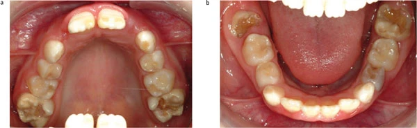 |
Difficulty brushing | Incisors and first permanent molars affected | |
Repeated restoration failure | Post-eruptive breakdown | |
Aesthetic concerns | Caries in affected molars |
Investigations
Primary investigations:
Full medical and dental history
Clinical examination and dental charting
Radiographs: bitewings to assess for interproximal caries, periapical radiographs if symptomatic