Clinical case
A 6-year-old child attends because the primary teeth are brown-blue, translucent and rapidly wearing down. The permanent incisors are beginning to erupt and appear similarly discoloured. Radiographs show bulbous crowns, cervical constriction and early obliteration of the pulp chambers. There is no history of bone fractures.
Pathophysiology
Dentinogenesis imperfecta results from abnormal dentine matrix formation and mineralisation. In non-syndromic cases, pathogenic variants in DSPP, which encodes dentine sialophosphoprotein, are commonly implicated. DSPP mutations are associated with dentinogenesis imperfecta types II and III and dentinal dysplasia type II.
The enamel is usually structurally normal, but it is poorly supported by abnormal dentine. This leads to chipping of enamel at the amelodentinal junction and exposure of defective dentine. The exposed dentine then wears rapidly.
Epidemiology and Risk Factors
Non-modifiable | Modifiable |
|---|---|
Family history | No known modifiable risk factors |
DSPP variant | |
Clinical Features
Symptoms | Signs | Radiographic signs | Image |
|---|---|---|---|
Discoloured teeth | Generalised involvement of many or all teeth | Bulbous crowns | 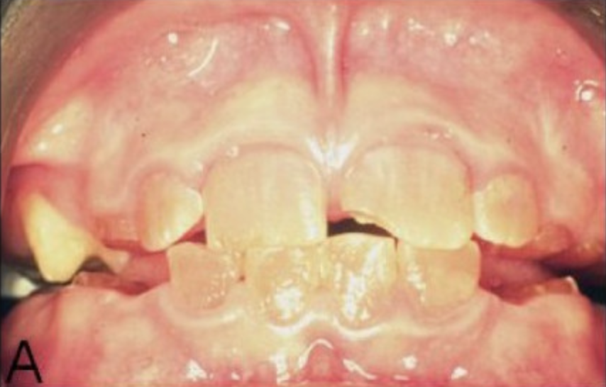 |
Rapid tooth wear | Primary dentition is worse affected | Cervical constriction | |
Sensitivity | Enamel chipping | Pulp chamber obliteration | |
Exposed soft dentine | 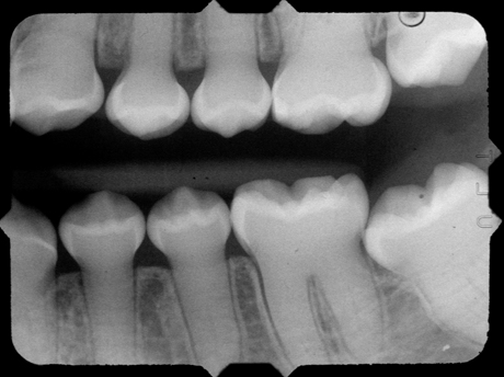 | ||
Reduced vertical dimension |
Investigations
Primary investigations:
Full medical and dental history
Clinical examination
Radiographs: OPG to assess roots and unerupted teeth, bitewings to assess for caries and pulp chamber changes, periapicals of affected teeth to assess roots and periapical status