The mandible is the only mobile bone of the maxillofacial skeleton. It forms the inferior border of the face and carries the lower teeth. Together with the maxillae, it forms the framework for the oral cavity and participates in mastication, speech and facial expression. It is a single bone in the adult, but develops from two halves that fuse in the midline during early childhood. The mandible articulates with the skull at the temporomandibular joints (TMJs) on each side, allowing complex movements of the lower jaw.
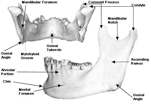
General Features
The mandible consists of:
A horizontal body (right and left parts, fused in the midline)
Two vertical rami (right and left)
The angle of the mandible at the junction between body and ramus
Each ramus bears two superior projections:
The coronoid process (anterior)
The condylar process (posterior), which ends in the head of the mandible that articulates with the temporal bone.
Body of the Mandible
The body is curved like a horseshoe and has external and internal surfaces, upper (alveolar) and lower (base) borders.
External Surface:
Feature | Characteristics |
|---|---|
Symphysis menti | A faint vertical ridge in the midline anteriorly, marking the side where the right and left halves of the mandible fused early in life |
Mental protuberance | A triangular bony prominence in the midline below the symphysis forming the chin |
Mental tubercles | Small elevations on either side of the mental protuberance |
Mental foramen | Located below the apices of the premolar teeth, this transmits the mental nerve and vessels which supply sensation to the lower lip and chin |
Oblique line | A ridge running upwards and backwards from the body of the mandible to the anterior ramus. It is a site of attachment for depressor anguli oris and depressor labii inferioris |
Internal Surface:
Feature | Characteristics |
|---|---|
Mental spines | Small bony projections near the midline
|
Mylohyoid line | A ridge running from behind the third molar region towards the symphysis area. It gives the origin for mylohyoid which forms the muscular floor of the mouth |
Sublingual foss | Above the anterior part of the mylohyoid line and houses the sublingual gland |
Submandibular foss | Below and behind the mylohyoid line and houses the submandibular gland |
Digastric foss | Depressions on the inferior border of the mandible near the midline; attachment for the anterior belly of digastric |
Alveolar Part
The alveolar process forms the upper border of the body and contains the sockets (alveoli) for the lower teeth.
In the dentate mandible, the alveolar process is prominent and contributes greatly to the height of the bone.
After tooth loss, the alveolar bone undergoes resorption, and the body appears much reduced in vertical height. This:
Alters the position of the mental foramen (often approaching the superior border).
Has major implications for complete denture construction and implant planning.
Ramus of the Mandible
Each ramus is a roughly quadrilateral plate of bone, extending upwards from the posterior part of the body.
Surface | Features |
|---|---|
Lateral surface | Provides attachment for masseter |
Medial surface |
|
The coronoid and condylar processes are located at the upper end of the ramus:
Coronoid Process
Thin, triangular and flattened.
Serves as the insertion for the temporalis muscle.
Lies beneath the zygomatic arch and moves within the temporal fossa during mandibular movements.
Condylar Process
The head (condyle) of the mandible – articulates with the articular disc and temporal bone in the TMJ.
The neck of the mandible – constricted region below the head, giving attachment anteromedially to part of the lateral pterygoid muscle.